

Young woman gets jabbed with a needle as a medical technician draws blood (Photo: US Navy)
A new device uses light instead of needles to test blood.
Researchers in Israel are developing the pain-free test, which is expected to reveal similar information as traditional blood tests, providing the doctor with immediate results.
Besides providing the patient with a relatively stress-free experience, the optical unit, which is no bigger than a breadbox, can also provide high-resolution images of blood flowing through our veins without the need for the harsh and short-lasting fluorescent dyes that are used in some test procedures.
“We have invented a new optical microscope that can see individual blood cells as they flow inside our body,” says Lior Golan, a graduate student at the Israel Institute of Technology, who co-authored an article in the Optical Society‘s journal Biomedical Optics Express.
While similar devices can “see” blood-flow, Golan says his team’s invention combines features which allow doctors to focus in on single cells, helping to provide additional information such as the shape and size of each cell, as well as other crucial diagnostic statistics.

An in vivo image shows red blood cells within a microvessel. (Photo: Biomedical Optics Express)
Other units are also far less practical, according to Golan, because they rely on bulky equipment and use potentially-harmful fluorescent dyes that must be introduced into the patient’s bloodstream.
The new microscope is based on a modest optical element and is driven by a technique called spectrally encoded confocal microscopy (SECM), which creates images by splitting a light beam into its basic colors -red, yellow, orange, green, cyan, blue, and violet- and emits them in a line of multi-colored light.
That rainbow-like light line is then focused across an appropriate blood vessel near the surface of the patient’s skin, and as flowing blood cells cross over the line it scatters the light.
The colors produced by the scattered light provide information which is interpreted by the system’s computer programs creating 2-D images of the blood cells.
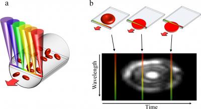
The team’s device relies on a technique called spectrally encoded confocal microscopy. (a) A single line within a blood vessel is imaged with multiple colors of light that encode lateral positions. (b) A single cell crossing the spectral line produces a two-dimensional image. (Photo: Biomedical Optics Express)
To use the device now, a doctor first attaches a small probe to the patient’s lower lip, with the patient’s blood vessels displayed on a nearby monitor.
After selecting a suitable vessel to help align and set the microscope, the doctor could begin to observe and analyze individual blood cells.
In the future, Golan says that his team is planning to miniaturize this device down to the size of a hand-held probe, devising a system that would automatically analyze the images, providing the doctor with valuable parameters and statistics to diagnose the patient.
Lior Golan joins us on this week’s radio edition of “Science World” to talk about how the device could one day become one your doctor’s most important diagnostic tools. Check out the right column for scheduled air-times or listen to the interview with Mr. Golan below.
[audio://blogs.voanews.com/science-world/files/2012/06/One-On-One-Lior-Golan-Needle-less-Blood-Tests.mp3|titles=One On One – Lior Golan – Needle-less Blood Tests]Other stories we cover on the “Science World” radio program this week include:
- SpaceX capsule Dragon returns from successful ISS mission
- Climate change might have sped collapse of ancient civilization
- Cell phones provide crucial health-information in Uganda
- Rare astronomical event ‘Transit of Venus‘ coming soon


is this out yet